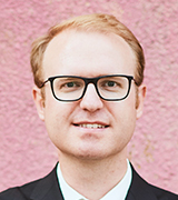
Chercheuse ou chercheur

    Benjamin De Leener , Ph.D. , Ing.

    benjamin.de-leener@polymtl.ca
    Benjamin De Leener
    Axe de recherche
    Santé musculosquelettique, réadaptation et technologies médicales
    Thème de recherche
    Maladies neurodéveloppementales

    Téléphone
    514-340-4711, poste 3689

    Sur le Web

    Titres

    Laboratoire

    Laboratoire de Recherche en Imagerie par Résonance Magnétique chez les enfants (laboratoire MAGIC)

    Formation

    • Postdoctorat, Institut de neurologie de Montréal, Université McGill, Montréal, Canada, 2018-2019
    • Postdoctorat, Département de génie électrique, Polytechnique Montréal, Montréal, Canada, 2017-2018
    • Ph.D., Génie biomédical, Polytechnique Montréal, Montréal, Canada, 2014-2017
    • M.Sc.A. Génie biomédical, Polytechnique Montréal, Montréal, Canada, 2012-2014
    • M.Sc.A., Ingénierie biomédicale, Université Libre de Bruxelles, Bruxelles, Belgique, 2011-2014
    • B. Ing., Ingénierie biomédicale, Université Libre de Bruxelles, Bruxelles, Belgique, 2007-2011

    Intérêts de recherche

    • Imagerie médicale
    • Vision par ordinateur
    • Neuroimagerie pédiatrique
    • Traitement et analyse d'images
    • Apprentissage machine
    • Modélisation

    Sujets CRSNG

    • 1901 Technologie biomédicale
    • 2603 Vision artificielle
    • 2605 Analyse de formes et intelligence artificielle
    • 2604 Applications de systèmes intelligents
    • 2708 Traitement d'images et traitement vidéo

    Sommaire de carrière

    Mes intérêts de recherche portent sur le développement de nouvelles technologies en neuroimagerie pédiatrique, avec un intérêt particulier sur les outils d'analyse d'images open-source du cerveau et de la moelle épinière acquises par résonance magnétique, en utilisant des approches avancées de segmentation et de recalage d'image, de template et d'atlas neurodéveloppementaux et d'apprentissage machine. En plus d’être situé à Polytechnique Montréal, le laboratoire MAGIC est lié à l'Institut TransMedTech et au Centre de Recherche du CHU Sainte-Justine, offrant des collaborations importantes entre chercheurs, ingénieurs, psychologue, radiologues et médecins.

    Prix et distinctions

    • Chaire TransMedTech en génie informatique, 2019-2021
    • Bourse d’études postdoctorales, HBHL, 2018-2019
    • Bourse d’études doctorales, FRQNT, 2016-2017
    • Étudiant chercheur étoile du FRQNT, 2016

    Financements majeurs

    • Chaire TransMedTech en génie informatique, 2019-2021

    Présentations

    • De Leener B, Solstrand Dahlberg L, Khatibi A, Cohen-Adad J, Doyon J. Effect of non-protonated perfluorocarbon liquid-filled SatPadsTM on spinal cord MR imaging. Proceedings of the 28th Annual Meeting of ISMRM, Sydney, Australia. 2020.
    • De Leener B, Solstrand Dahlberg L, Khatibi A, Kinany N, Doyon J. Improving spatial normalization of functional MRI data of the spinal cord using cerebrospinal fluid segmentation. Proceedings of the 28th Annual Meeting of ISMRM, Sydney, Australia. 2020.
    • Ouellette R, Treaba CA, Granberg T, Barletta V, Herranz E, Mangeat G, et al. Characterization of multiple sclerosis grey and white matter pathology in the brain and spinal cord at 7 Tesla MRI. ECTRIMS 2018. 2018.
    • Shahrampour S, De Leener B, Middelton D, Flanders A, Faro S, Cohen-Adad J, et al. Volumetric Analysis of Pediatric Spinal Cord Structural MRI data using SCT. Proceedings of the 56th Annual Meeting and the ASNR Symposium 2018. 2018. p. O433.
    • De Leener B, Fonov V, Collins DL, Callot V, Stikov N, Cohen-Adad J. PAM50: Multimodal template of the brainstem and spinal cord compatible with the ICBM152 space. Proceedings of the 25th Annual Meeting of ISMRM, Honolulu, USA. 2017.

    Publications

    Consulter la liste complète.

    • Eden D, Gros C, Badji A, Dupont SM, De Leener B, Maranzano J, et al. Spatial distribution of multiple sclerosis lesions in the cervical spinal cord. Brain. 2019;142: 633–646. doi:10.1093/brain/awy352
    • Gros C, De Leener B, Badji A, Maranzano J, Eden D, Dupont SM, et al. Automatic segmentation of the spinal cord and intramedullary multiple sclerosis lesions with convolutional neural networks. Neuroimage. arxiv.org; 2019;184: 901–915. doi:10.1016/j.neuroimage.2018.09.081
    • Martin AR, De Leener B, Cohen-Adad J, Kalsi-Ryan S, Cadotte DW, Wilson JR, et al. Monitoring for myelopathic progression with multiparametric quantitative MRI. PLoS One. 2018;13: e0195733. doi:10.1371/journal.pone.0195733
    • De Leener B, Fonov VS, Collins DL, Callot V, Stikov N, Cohen-Adad J. PAM50: Unbiased multimodal template of the brainstem and spinal cord aligned with the ICBM152 space. Neuroimage. 2018;165: 170–179. doi:10.1016/j.neuroimage.2017.10.041
    • Gros C, De Leener B, Dupont SM, Martin AR, Fehlings MG, Bakshi R, et al. Automatic spinal cord localization, robust to MRI contrasts using global curve optimization. Med Image Anal. 2017;44: 215–227. doi:10.1016/j.media.2017.12.001
    • Prados F, Ashburner J, Blaiotta C, Brosch T, Carballido-Gamio J, Cardoso MJ, et al. Spinal cord grey matter segmentation challenge. Neuroimage. 2017;152: 312–329. doi:10.1016/j.neuroimage.2017.03.010
    • De Leener B, Mangeat G, Dupont S, Martin AR, Callot V, Stikov N, et al. Topologically preserving straightening of spinal cord MRI. J Magn Reson Imaging. 2017; doi:10.1002/jmri.25622
    • Martin AR, De Leener B, Cohen-Adad J, Cadotte DW, Kalsi-Ryan S, Lange SF, et al. Clinically Feasible Microstructural MRI to Quantify Cervical Spinal Cord Tissue Injury Using DTI, MT, and T2*-Weighted Imaging: Assessment of Normative Data and Reliability. AJNR Am J Neuroradiol. 2017;38: 1257–1265. doi:10.3174/ajnr.A5163
    • Martin AR, De Leener B, Cohen-Adad J, Cadotte DW, Kalsi-Ryan S, Lange SF, et al. A Novel MRI Biomarker of Spinal Cord White Matter Injury: T2*-Weighted White Matter to Gray Matter Signal Intensity Ratio. AJNR Am J Neuroradiol. 2017;38: 1266–1273. doi:10.3174/ajnr.A5162
    • De Leener B, Lévy S, Dupont SM, Fonov VS, Stikov N, Louis Collins D, et al. SCT: Spinal Cord Toolbox, an open-source software for processing spinal cord MRI data. Neuroimage. 2017;145: 24–43. doi:10.1016/j.neuroimage.2016.10.009
 

Voir Grand pour l’amour des enfants

Avec le soutien de donateurs comme vous au cœur de la campagne majeure Voir Grand, nous conduisons les équipes soignantes vers les opportunités de la science et des nouvelles technologies pour que chaque enfant, où qu’il soit au Québec, accède au savoir-faire et au savoir-être uniques du CHU Sainte-Justine. Ensemble, unissons nos forces pour leur avenir.

Merci de voir grand avec nous.

Vous pouvez également faire votre don par la poste ou par téléphone au numéro sans frais 1-888-235-DONS (3667)

Nous contacter

514 345-4931

Légal

© 2006-2014 Centre de recherche Azrieli du CHU Sainte-Justine.
Tous droits réservés.
Avis légaux  Confidentialité  Sécurité

Avertissement

Les informations contenues dans le site « CHU Sainte-Justine » ne doivent pas être utilisées comme un substitut aux conseils d’un médecin dûment qualifié et autorisé ou d’un autre professionnel de la santé. Les informations fournies ici le sont à des fins exclusivement éducatives et informatives.

Consultez votre médecin si vous croyez être malade ou composez le 911 pour toute urgence médicale.

CHU Sainte-Justine